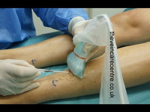
An essential skill in modern phlebology is the ability to cannulate blood vessels under ultrasound guidance. They can be can be cannulated in transverse section or longitudinal section and both methods have their advocates. But which is best? Well I am going to go through the pros and cons of each method show you each method being performed and then I am going to tell you my own preference. This schematic shows the appearance of the vessel when it is imaged in transverse section. Here we can see the vessel running longitudinally and the ultrasound probe is placed at 90° to it so that the long axis of the transducer is at 90° to the long axis of the blood vessel. The image that is produced is therefore a circle. When the blood vessel is imaged in longitudinal section the long axis of the ultrasound probe is parallel and directly over the long axis of the blood vessel. And in this situation the ultrasound image looks like this: 2 parallel lines indicating the superficial and deep walls of the blood vessel and the lumen in between. In this schematic diagram we can see the cannulation being performed in longitudinal section.
The ultrasound probe has its long axis parallel to the long axis of the blood vessel and directly over it and the needle is being inserted at an angle directly over the blood vessel. In longitudinal section this is the ultrasound appearance: the superficial and deep walls of the blood vessel are seen and the entire length of the needle is seen on the ultrasound as it passes from superficial to deep. Here we can see the blood vessel being cannulated with the transducer in transverse section. The long axis of the transducer is at 90° to the long axis of the blood vessel and the probe is here directly over the needle tip as it is entering the blood vessel. On the ultrasound we can see the blood vessel as a circle and the tip of the needle appears as an echogenic point within the lumen of the blood vessel. Here we can see the vein being imaged in transverse section and compressed with the probe. Here is the corresponding ultrasound appearance. Here the probe is rotated at 90° and the vein is imaged in longitudinal section. Here is the corresponding image. The vein can be cannulated under transverse section and here we can see the tip of the needle coming down into the lumen of the vein and in longitudinal section we can see the needle being advanced within the lumen of the blood vessel under ultrasound guidance.
So what are the pros and cons of cannulation in longitudinal section? Well 1st of all with the probe directly over the blood vessel and the long axis of the probe in the long axis of the blood vessel we can on the ultrasound see the whole length of the needle as it advances through the superficial wall of the vein and into the lumen. We can keep the probe absolutely still and the only movement is the movement of the needle going into the blood vessel. The disadvantages are that of course you only see the superficial and deep walls of the blood vessel. It can sometimes be very difficult to keep the ultrasound probe directly over the blood vessel and directly parallel in the same plane. And for these reasons small blood vessels small veins can be very challenging certainly in my experience of using this method. What about the pros and cons of cannulation in transverse section? Well here you can see a schematic of the needle entering the vein and in fact making its way through the vein and through the deep wall of the vein.
The ultrasound probe has to be moved and these are the positions I’ve chosen to show you: point A where the vein is punctured point B where the needle has gone into the lumen and points C where the needle has gone through the blood vessel and come out the back. Now let’s assume that the needle has gone all the way through and out the back if you image the vein at point A you see the needle apparently within the lumen just entering the lumen and that would be the case as the needle was advanced. There it is the echogenic tip in the lumen and at C you can see needle has gone through the back. Now the advantages of this transverse method is that you see the entire circumference of the vein so you see not only the superficial wall and the deep wall but you see the lateral and medial walls as well.
And for this reason certainly my experience is that transverse imaging does facilitate the cannulation of very small veins. The disadvantage of the method is that if you keep the transducer stationary you of course don’t see the whole length of the vessel you have to move the transducer as the needle advances. So there is movement of the probe as well as movement of the needle. You need to coordinate movement of the probe with the advancement of the needle. But if you are able to coordinate this movement of the transducer as the need advances I think you you can better and more easily cannulate very small blood vessels but you do need very good imaging to see the needle tip.
So there you have the pros and cons of each method. You’ve seen each method being performed. Which is my preference? Well my preference is to perform cannulation almost exclusively under transverse imaging. I find that I can cannulate very small blood vessels easily and I’ve got used to coordinating movements of the probe with my left hand moving it up and down the length of the blood vessel whilst moving the needle with my right hand. And in this way I can build up a 3-D image in my own mind about the course of the blood vessel I can picture where the needle tip is in relation to the blood vessel and I can cannulate blood vessels 1 to 2 mm in diameter. So there you have the pros and cons of longitudinal and transverse imaging. I think in summary it is fair to say that it is a personal choice. I don’t think there is a right or a wrong way. I would recommend that you try both see which suits you and if you are cannulating blood vessels successfully with either method then that’s the one for you.
As found on Youtube
