– [Voiceover] A linear array transducer with a venous exam type, is used to perform an ultrasound guided insertion of a subclavian vein catheter via an transverse approach. The patient is in a supine position with the head neutral. The operator should stand to the patient’s right side. The transducer is placed transversely just inferior to the mid portion of the clavicle, with the orientation marker directed to the patient’s head at a 12 o’clock position.
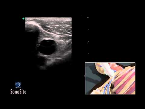
The hyperechoic clavicle can be seen in the superior portion of the ultrasound image. The vein is dark, and anechoic, just inferior and deep to the clavicle. The transducer should be slowly moved one to two inches toward the shoulder, with the face of the transducer staying below the clavicle to obtain the best view of the subclavian or axillary vein. It is important to note that the lung lies directly posterior to the vessel. So, posterior wall puncture of the axillary vein should be avoided. Adjust the transducer so it is centered over the vein. Follow the needle entry by slowly sliding the transducer in the direction of needle advancement. The needle will appear as a small, bright, hyperechoic dot. When the needle tip appears, the transducer should be advanced a short distance distally to follow the tip of the needle trajectory, and stay in advance of the needle entry.
The needle is slowly advanced under direct ultrasound visualization until the tip is seen to puncture the subclavian vein. The probe should be moved slightly proximally and distally to confirm that the needle tip lies in the mid portion of the vein..