[Radiologist] So this is just a view of a liver. [Kevin Parker] For years people have been trying to improve ultrasound. [Radiologist] So there should be something right up here and we’re not seeing it very well. [Kevin Parker] When you are in the solid tissue of like the liver or the thyroid or the breast, it’s really hard to distinguish what’s going on inside there. [Deborah Rubens] So ultrasound began forty, fifty years ago and it was originally developed to look at babies in utero and from there it’s grown to a tremendous tool that can examine almost any kind of soft tissue.
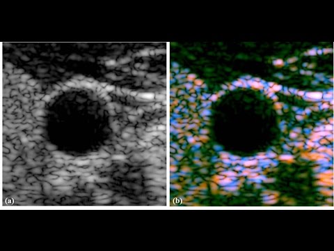
Ultrasound energy, when it hits a surface, it doesn’t just sort of reflect straight back necessarily and gives us a picture sometimes the energy goes in different directions and scatters and so it’s not a perfect image. [Kevin Parker] So on the outside of the screen is the old fashioned black and white B-scan. What we’ve done here is sort of a very special analysis based on what comes back from larger and smaller objects. They have a certain signature and we’re looking for that signature mathematically and then we display it as colors and the color tells you something about what the scatters are inside the tissue, inside the cells. The doctors are always looking at livers and thyroids and breast tissue and others and they might sight see some unusual areas so that’s a lesion. Well it would be great if we could just characterize that tissue and know just by the scan and just by the colors and be reassured that it’s normal or benign, basically.
So, in many cases a normal tissue will be somewhat greenish and then the unusual things will show up as bright blue spots or orange-red spots. [Deborah Rubens] On thing that happens that we’re involved with in medicine is tumors and tumor therapy so if you could look at something and figure out is the therapy working, that would be useful. [Kevin Parker] Ultrasound is for examples going into miniaturized versions that people can hold in their pockets and take out to rural clinics, for example, so it has a lot of uses, not just in the big hospitals but sort of out in clinics everywhere, even in the developing world. [Deborah Rubens] Sometimes replacing maybe a CT scan or an MR scan where we might have that information but ultrasound can do it cheaper, better, faster, at the bedside, at the place where you’re going to do your intervention. [Kevin Parker] At the end of the day if it really makes an improvement then it starts to disseminate worldwide.
It’s a very nice pathway but it is one that takes years and years of work to get it disseminated..
As found on Youtube